POTS Blood Tests and Autonomic Lab Work
Different blood markers identify which of three distinct mechanisms is driving a patient's POTS.

POTS blood work exists to answer one question: which of several different physiological problems is causing this patient's heart rate to spike when they stand up. That question has more than one right answer. The testing process looks less like a single checklist and more like a branching investigation as a result.
Why POTS Is Not a Single Disease
Postural orthostatic tachycardia syndrome is defined by a pattern: a heart rate increase of 30 beats per minute or more in adults, 40 or more in adolescents, within minutes of standing, without the blood pressure drop that marks orthostatic hypotension, once other causes have been excluded. That definition describes what the body does. It says nothing about why the body does it, and the "why" turns out to split into at least three distinct mechanisms.
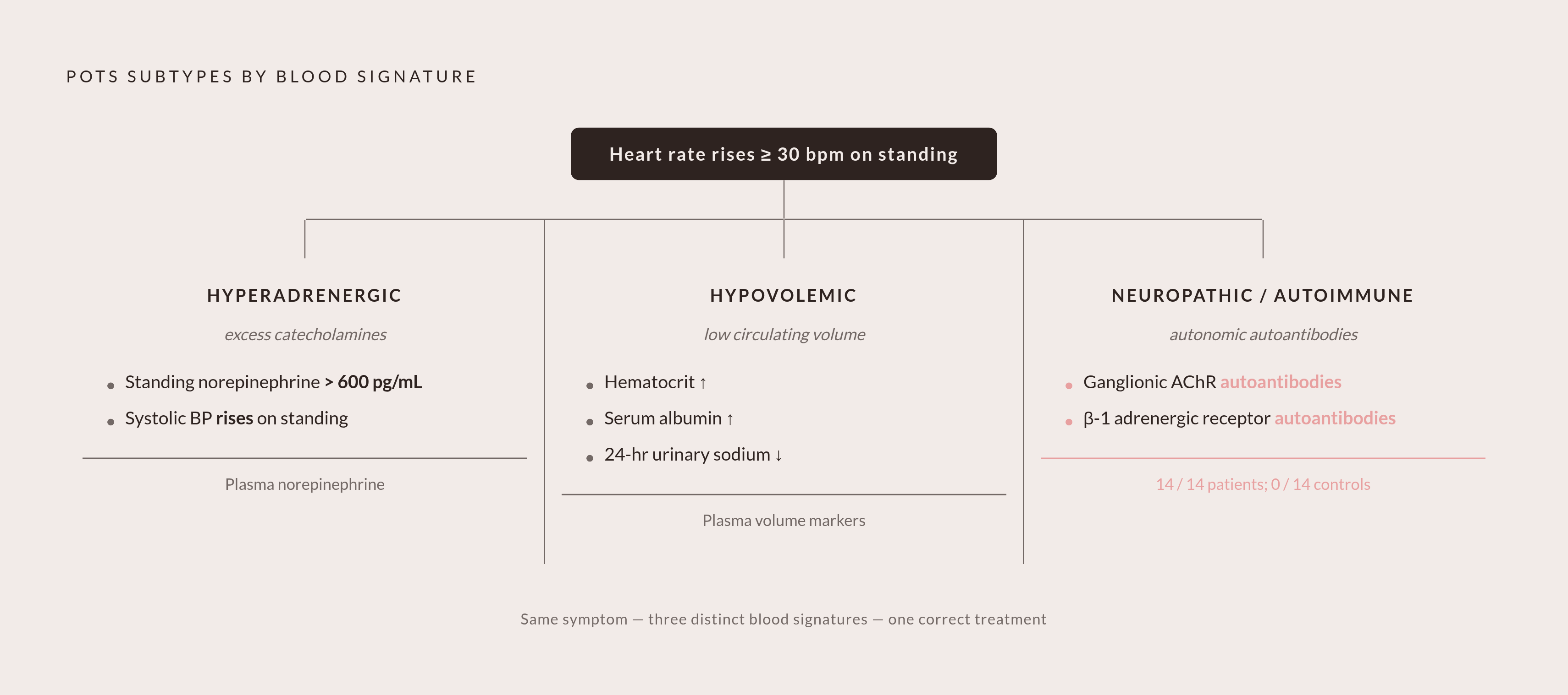
Some patients develop POTS because their sympathetic nervous system overreacts to standing, flooding the body with norepinephrine. Others develop it because they simply don't have enough blood volume circulating, so the heart compensates by beating faster when gravity pulls blood toward the legs. Still others develop it because of nerve or receptor damage, often autoimmune in origin, that interferes with normal blood vessel constriction. These are called the hyperadrenergic, hypovolemic, and neuropathic or autoimmune subtypes, and each one leaves a different signature in the blood.
The population affected by this condition has grown substantially in recent years. A 2025 study in Heart Rhythm O2 put POTS prevalence at 3.4% following COVID-19 infection, naming it one of the most common forms of cardiovascular autonomic dysfunction that follows the virus. That means a large and growing number of patients are entering a diagnostic process that was already complicated before long COVID made it more urgent.
The practical consequence for anyone reading their own lab results: a number that looks normal for one subtype can be the exact abnormality that confirms another. A clinician who doesn't know which mechanism they're investigating can't interpret a single value correctly, and neither can a patient trying to make sense of a report handed across a desk. The workup that follows is a structured test of competing explanations, where each result either rules a mechanism in or rules it out, not a routine panel run out of habit.
The tests that rule out other causes before POTS can be confirmed
The first round of tests must clear away conditions that look like POTS but aren't before any test can point toward a POTS subtype. That's the real job of the first round of blood work: eliminating other explanations, not confirming this one. A normal result at this stage is the necessary first step.
The standard exclusionary panel checks for anemia through a complete blood count, electrolyte imbalances that can throw off autonomic regulation, and thyroid dysfunction, which produces the same racing heart and fatigue that POTS does. These conditions are common and treatable, so no clinician should move toward autonomic-specific testing without ruling them out first.
Serum albumin often gets checked alongside these, since low albumin can contribute to plasma volume problems and shows up in formal POTS protocols. A 24-hour urinary sodium test, while not a blood draw, frequently accompanies this panel: low sodium excretion signals that a patient isn't taking in enough salt, which directly affects how much plasma volume they're carrying and shapes whether salt supplementation makes sense as a next step.
Here is where the diagnostic process tends to go wrong for a lot of patients. When these exclusionary tests come back normal, a clinician unfamiliar with POTS may read that as "there's nothing wrong," instead of understanding that normal exclusionary results are where the real investigation into autonomic dysfunction begins. The Australian POTS Patient Registry study found that 64.6% of patients had their symptoms attributed to anxiety before they were diagnosed, a figure that lines up closely with what happens when a clinician stops looking after a normal basic panel instead of continuing toward the tests that actually probe autonomic function. The frustration patients feel in that moment makes sense. The bloods aren't lying. The bloods are answering the question clinicians keep asking instead of the one that matters.
What plasma norepinephrine levels reveal about hyperadrenergic activity
Once the exclusionary panel is clear, testing moves from "what else could this be" to "which mechanism is this." The first mechanism-specific test measures plasma norepinephrine, and it's checking whether the sympathetic nervous system is overactivating when a patient stands, which is the defining trait of hyperadrenergic POTS.
Position and timing matter here because blood gets drawn twice, once while the patient is lying flat, usually after an extended period of rest or overnight, and again after they've stood up. Drawing twice captures the change between the two positions, because that change is the actual signal a clinician is looking for, not redundancy. A single norepinephrine reading taken in isolation tells almost nothing.
A standing norepinephrine level above 600 pg/mL supports a hyperadrenergic pattern, especially when paired with a rise in systolic blood pressure on standing rather than the drop seen in orthostatic hypotension. That combination indicates the sympathetic nervous system is dumping excess norepinephrine into circulation in response to upright posture, and that surge is what's driving the heart rate spike.
The opposite pattern matters too. In some autonomic neuropathies, called pandysautonomia, norepinephrine levels stay low even on standing and fail to rise at all. That's a different kind of autonomic failure entirely, one that points toward peripheral nerve dysfunction rather than sympathetic overactivation. Getting this distinction right shapes which treatments make sense, since beta-blockers and other drugs that dampen sympathetic activity work differently depending on whether norepinephrine is elevated or depleted. Treating the wrong subtype can make a patient's symptoms worse rather than better.
One more step belongs in this part of the workup. A rare adrenal tumor called pheochromocytoma produces norepinephrine and can mimic hyperadrenergic POTS closely enough that plasma metanephrine testing is used to rule it out before a hyperadrenergic diagnosis is confirmed. Pheochromocytoma itself is uncommon, but the two conditions can look alike on paper, and a clinician needs to know which one is actually present before building a treatment plan.
How blood volume testing identifies the hypovolemic subtype
A different mechanism requires a different kind of test. Plasma volume deficit appears in markers of how much fluid is actually circulating in the body rather than in a hormone reading, and a large share of POTS patients carry a measurable shortfall here, one that directly shapes whether increased fluid and salt intake makes sense as a first-line approach.
The mechanism is mechanical rather than hormonal. When there isn't enough circulating blood volume, less blood returns to the heart once a patient stands up, so the heart compensates by beating faster. The resulting symptom looks identical to hyperadrenergic POTS from the outside, but the cause is a volume problem rather than a sympathetic nervous system overreacting to posture. That's precisely why this workup can't rely on heart rate and symptoms alone to pick a treatment path.
Standard labs can hint at this deficit without measuring it directly. Hematocrit and red blood cell indices from a complete blood count can suggest hemoconcentration, which is a marker of low plasma volume, and serum albumin adds another layer of signal. For a more exact measurement, researchers use a technique called CO rebreathing, which quantifies plasma volume and red blood cell mass directly. This method is being used in an NHLBI-funded observational study led by Dr. Steven Vernino at UT Southwestern, which is phenotyping POTS patients in depth using this technique alongside immunophenotyping and neurohormonal assessments. That a method this precise is being built into a major research protocol shows plasma volume is treated as a measurable, quantifiable variable.
The 24-hour urinary sodium test connects directly to this picture. Low sodium excretion means the kidneys are holding onto sodium aggressively, which is the body's way of compensating for low volume, and the same test also tells a clinician whether a patient's salt intake is adequate to support treatment. For patients, this has a practical use beyond the lab report itself: demonstrating a measurable plasma volume deficit gives real grounding to requests for evidence-based treatments like salt loading, structured fluid protocols, or compression garments, and it explains clearly why generic advice to "just drink more water" misses what's actually happening in the body.
What autoimmune markers are revealing about nerve and receptor damage
The third mechanism is also the one where the science is moving fastest. A growing body of evidence points to an autoimmune process in a meaningful subset of POTS patients, with specific autoantibodies, particularly those targeting adrenergic and muscarinic receptors, under active study as potential diagnostic markers.
In neuropathic POTS, blood pools excessively in the arms and legs when a patient stands because the peripheral nerves responsible for constricting blood vessels aren't doing their job. One proposed cause is autoimmune damage to those nerves or the receptors they depend on. Several specific antibodies are under investigation. Ganglionic acetylcholine receptor autoantibodies have been linked to autoimmune autonomic neuropathy and found in some neuropathic POTS patients, targeting the nerve ganglia that regulate the whole autonomic system. Adrenergic receptor autoantibodies targeting alpha-1, beta-1, and beta-2 receptors have shown an even more striking finding in one study: serum from all 14 POTS patients tested produced dose-dependent activation of beta-1 adrenergic receptors, compared to none of the healthy controls tested.
These findings matter, but none of these autoantibody tests are part of standard clinical panels in most healthcare systems. They're available mainly through specialist centers and research protocols, so a patient whose workup doesn't include them hasn't necessarily been tested poorly, just not tested at the current research frontier. The NHLBI-funded phenotyping study at UT Southwestern, registered as NCT06292104 on ClinicalTrials.gov, includes immunophenotyping as a core piece of its design, reflecting a broader shift in the field toward treating autoimmunity as a primary target of investigation rather than an afterthought. That same protocol includes a skin biopsy measuring intraepidermal nerve fiber density, which checks for small fiber neuropathy directly at the tissue level and backs up what the blood-based antibody testing suggests. Between the two, the investigation into this subtype is reaching past blood alone and into tissue itself, a sign of how seriously researchers are chasing down the autoimmune thread.
Functional Tests and Lab Work in the Diagnostic Picture
None of the blood tests described above mean much read in isolation. They're interpretable because they're paired with functional tests that capture what actually happens to a patient's body in real time when they move from lying down to standing up.
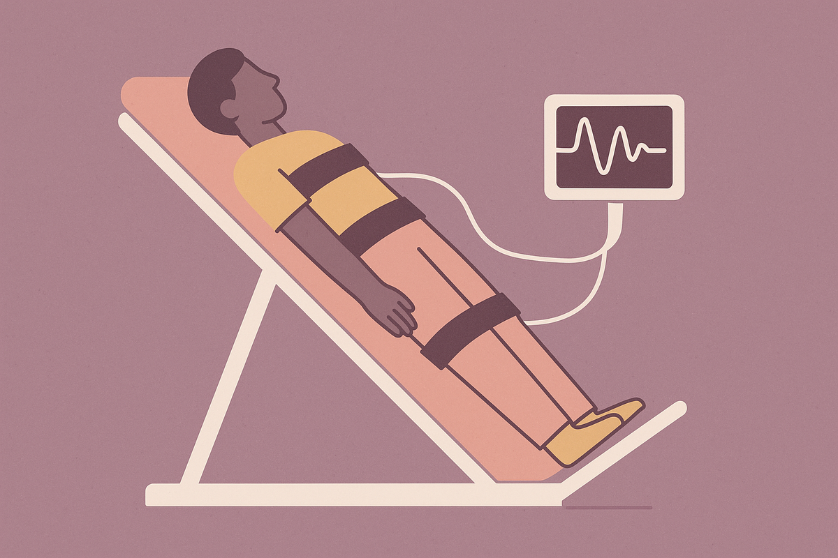
The tilt table test is the most controlled version of this. A patient lies on a table that tilts gradually upright to simulate standing, while continuous monitoring tracks heart rate and blood pressure throughout. A positive result for POTS requires a sustained heart rate increase of at least 30 beats per minute in adults, or 40 in adolescents, with blood pressure holding steady rather than dropping. The active standing test offers a more accessible alternative for general practice settings: heart rate and blood pressure are recorded while the patient lies down, then again after ten minutes of standing. It's less controlled than a tilt table, but the same diagnostic threshold applies.
Position also matters this much for the blood tests described earlier. A norepinephrine level drawn only while supine, or only while standing, loses most of its diagnostic value, since the change between positions carries the signal rather than either number alone.
The NASA Lean Test deserves a fair and careful word here, because many patients have used it to flag their own symptoms before ever reaching a clinician. A 2025 paper by Teuschl and colleagues in Clinical Autonomic Research found that despite how widely the NASA Lean Test gets used, validation studies comparing its diagnostic accuracy against tilt table testing or the active standing test are lacking. The formal diagnostic criteria for POTS and orthostatic hypotension rest on hemodynamic data from head-up tilt or active standing tests, not lean tests, and self-diagnosis based on a lean test alone has carried a high rate of inaccuracy. None of that makes the test useless. It has a real role in flagging who might need a closer look. The shortfall sits with healthcare systems that too often stop at a patient's self-reported lean test result instead of following up with one of the formal assessments that can actually confirm or rule out POTS.
A related tool is showing promise for exactly that kind of triage. Hupin and colleagues, writing in Heart Rhythm O2 in 2025, tested 24-hour ECG recording in long-COVID patients, analyzing heart rate spikes, the rise in heart rate on waking, and heart rate variability, as a way to direct clinical suspicion toward POTS before formal autonomic testing. Used this way, a day-long ECG becomes a bridge, helping decide who needs to be referred for tilt table or active standing testing rather than serving as a diagnosis on its own.
Put together, blood work and functional testing can establish which subtype a patient has, how severe it is, which treatment targets make the most sense, and, just as important for the patient sitting across the desk, a documented and objective record of what their autonomic nervous system is actually doing.
Why Diagnostic Delays Persist
Every test described in this piece exists today. Most patients still spend years reaching them, held back by clinicians unfamiliar with POTS, by symptoms wrongly attributed to anxiety, and by referral pathways that don't route patients toward the right specialists in time.
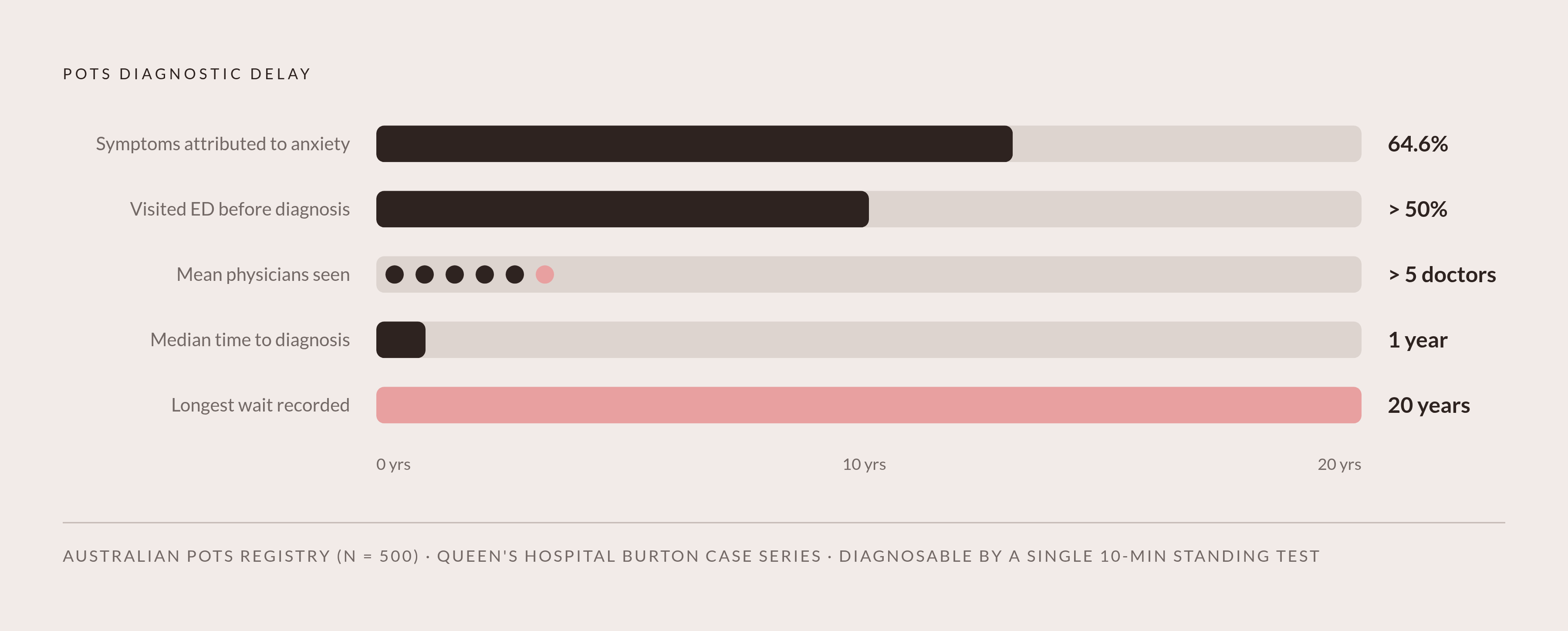
The Australian POTS Patient Registry, drawing on 500 clinically diagnosed patients, found that more than half had been to the emergency department for POTS-related symptoms before ever being diagnosed. Almost two-thirds had their symptoms attributed to anxiety at some point along the way. Patients saw a mean of more than five physicians before getting a diagnosis, and the delay stretched longer for women than for men.
A case series from Queen's Hospital Burton, a single-center UK district general hospital, found a median time to diagnosis of one year, with delays stretching as long as 20 years for some patients in that same setting. A one-year median sounds almost reasonable next to the years-long ordeals that show up in patient registries, until you remember that for a condition defined by a 10-minute standing test, a year is still an extraordinarily long wait to confirm something measurable in a single clinic visit.
None of this reflects a shortage of tools. The norepinephrine draw, the plasma volume markers, the tilt table test, the autoantibody panels moving through research centers: every piece of this diagnostic architecture already exists and already works. What's missing more often is a clinician who recognizes POTS early enough to order the right test in the right sequence, and a referral system that gets a patient to that clinician before five other doctors and an emergency room visit come first.
Sources
- Phenotyping of Postural Orthostatic Tachycardia Syndrome (POTS)
- Symptom burden, quality of life, and diagnostic journey of people with postural orthostatic tachycardia syndrome, Australia, 2021–24: a descriptive patient registry data study
- Is the NASA Lean Test a suitable tool to diagnose cardiovascular autonomic disorders?
- Clinical Presentation, Diagnostic Delays, and Treatment Outcomes in Postural Orthostatic Tachycardia Syndrome (POTS): An Observational Case Series Study in a Single-Centre District General Hospital
- Screening for postural orthostatic tachycardia syndrome using 24-hour electrocardiogram recording in patients with long coronavirus disease
- POTS Diagnostic Criteria: How Postural Orthostatic Tachycardia Is Diagnosed
- Postural orthostatic tachycardia syndrome: insights into pathogenesis and treatment
- Blood volume deficit in postural orthostatic tachycardia syndrome assessed by semiautomated carbon monoxide rebreathing - PMC